March 28, 2016
Ailments
What is tennis elbow and what are the symptoms? Tennis elbow causes pain on the outer side of your elbow. The medical term for tennis elbow is lateral epicondylitis. This is because the pain is felt around the area of the lateral epicondyle (the lower, outer, bumpy part of your humerus bone in your upper arm).For most people with tennis elbow, the pain only occurs when they use their forearm and wrist, particularly for twisting movements such as turning a door handle or opening a jar. However, for some people the pain is constant; it occurs at rest and can affect their sleep. The pain may travel down your arm from your elbow towards your wrist. You may find it difficult to hold items such as a knife or fork, a cup or a pen, or to straighten your arm fully. Some people also notice a stiffness in the affected arm.Golfer's elbow is the name given to a similar condition that produces pain around the inner side of your elbow.

Image from google images What is a prolapsed disc?A 'slipped' (prolapsed) disc often causes severe lower back pain. The disc often presses on a nerve root which can cause pain and other symptoms in a leg. In most cases, the symptoms ease off gradually over several weeks. The usual advice is to do normal activities as much as possible. Painkillers may help. Physical treatments such as spinal manipulation may also help. Surgery may be an option if the symptoms persist.

When you have a 'slipped' (prolapsed) disc, a disc does not actually slip. What happens is that part of the inner softer part of the disc (the nucleus pulposus) bulges out (herniates) through a weakness in the outer part of the disc. A prolapsed disc is sometimes called a herniated disc. The bulging disc may press on nearby structures such as a nerve coming from the spinal cord. Some inflammation also develops around the prolapsed part of the disc.Any disc in the spine can prolapse. However, most prolapsed discs occur in the lower back (the lumbar spine). The size of the prolapse can vary. As a rule, the larger the prolapse, the more severe the symptoms are likely to be.What are the types of low back pain?Nonspecific low back painThis is the most common type of back pain. The majority of cases of sudden-onset (acute) low back pain are classed as nonspecific. This is the type of back pain that most people will have at some point in their life. It is called nonspecific because it is usually not clear what is actually causing the pain. In other words, there is no specific problem or disease that can be identified as the cause of the pain. The severity of the pain can vary from mild to severe. This type of back pain is discussed further below.Nerve root pain - often called sciaticaThis occurs in less than 1 in 20 cases of acute low back pain. Nerve root pain means that a nerve coming out from the spinal cord (the root of the nerve) is irritated or pressed on. (Many people call this a trapped nerve.) You feel pain along the course of the nerve. Therefore, you typically feel pain down a leg, sometimes as far as to the calf or foot. The pain in the leg or foot is often worse than the pain in the back. The irritation or pressure on the nerve may also cause pins and needles, numbness or weakness in part of a buttock, leg or foot.About 9 in 10 cases of nerve root back pain are due to a prolapsed disc - often called a slipped disc. (A disc does not actually slip. What happens is that part of the inner softer part of the disc bulges out (prolapses) through a weakness in the outer harder part of the disc. The prolapsed part of the disc can press on a nerve nearby. See separate leaflet called Prolapsed Disc (Slipped Disc) for details.) Other less common conditions can cause pressure on a nerve to cause nerve root pain.Frozen ShoulderFrozen shoulder (sometimes called adhesive capsulitis of the shoulder) is a condition where a shoulder becomes painful and stiff. Shoulder movements become reduced, sometimes completely 'frozen'. It is thought to be due to scar-like tissue forming in the shoulder capsule. Without treatment, symptoms usually go but this may take up to 2-3 years. Various treatments may ease pain and improve the movement of the shoulder.What are the symptoms of frozen shoulder?The typical symptoms are pain, stiffness, and limitation in the range of movement of a shoulder. The symptoms typically have three phases:
- Phase one - the 'freezing', painful phase. This typically lasts 2-9 months. The first symptom is usually pain. Stiffness and limitation in movement then also gradually build up. The pain is typically worse at night and when you lie on the affected side.
- Phase two - the 'frozen', stiff (or adhesive) phase. This typically lasts 4-12 months. Pain gradually eases but stiffness and limitation in movement remain and can get worse. All movements of the shoulder are affected. However, the movement most severely affected is usually rotation of the arm outwards. The muscles around the shoulder may waste a bit as they are not used.
- Phase three - the 'thawing', recovery phase. This typically lasts between one and three years. The pain and stiffness gradually go and movement gradually returns to normal, or near normal.
Symptoms often interfere with everyday tasks such as driving, dressing, or sleeping. Even scratching your back, or putting your hand in a rear pocket, may become impossible. Work may be affected in some cases.There is great variation in the severity and length of symptoms. Untreated, on average the symptoms last 2-3 years in total before going. In some cases, it is much less than this. In a minority of cases, symptoms last for several years.What is the carpal tunnel?There are eight small bones called carpal bones in the wrist. A ligament (also called retinaculum) lies across the front of the wrist. Between this ligament and the carpal bones is a space called the carpal tunnel. The tendons that attach the forearm muscles to the fingers pass through the carpal tunnel. A main nerve to the hand (median nerve) also goes through this tunnel before dividing into smaller branches in the palm.The median nerve gives feeling to the thumb, index and middle fingers, and half of the ring finger. It also controls the movement of the small muscles at the base of the thumb.

What is carpal tunnel syndrome?This syndrome is a set of symptoms thought to be caused by squashing (compression) of the median nerve in the carpal tunnel. In terms of age, carpal tunnel syndrome is more common in:
- People in their late 50s, particularly women.
- People in their late 70s, when men and women are equally affected.
Carpal tunnel syndrome is more common in people who are obese and it often runs in families. It is more common in women who are pregnant.Causes of back painNon-specific - the cause in the vast majority of peopleIt is often impossible to find a precise cause for low back pain. Less than 1 in 100 people have a serious problem. It can be caused by an injury or sprain, but most of the time it isn't and may be due to poor posture, lack of exercise or stiffness. You may have heard your doctor, physiotherapist or nurse describing your back pain as 'non-specific' or 'simple' back pain. This means that after your examination, the clinician is not concerned that you have a serious medical condition. This is the type of back pain that is likely to get better over the next few weeks as you gradually return to normal activities and work.SciaticaThis is far less common and affects less than 1 in 20 people. It is most often caused by pressure or irritation of nerves as they come out of the lower back. The symptoms include pain, numbness and tingling that spread down the leg, sometimes reaching the calf or foot. Most people do recover from sciatica over time but often it takes longer than with non-specific back pain.ArthritisArthritisis common. There are many different causes of arthritis and so all agegroups can be affected. Some causes of arthritis only cause joint problems for ashort time and there are no long-lasting problems. Other causes of arthritis maycause pain and difficulties for a long period of time or even permanently.If you have any joint problems you should see your GP to find out the cause of thearthritis. You will often need some investigations, such as blood tests and X-rays.You may also need to be seen by a specialist, depending on the likely cause of arthritisWhat is arthritis?Arthritis means inflammation of joints. Arthritis is very common. There are many different causes of arthritis. Children and adults of all ages can be affected by arthritis.Arthritis may affect just one joint, a few joints or many joints. Each cause of arthritis tends to have a typical pattern in terms of which joints are affected and the age of people most likely to be affected.In the knee, there are areas of cartilage tissue which act like shock absorbers in the joint - these are called menisci. There are also areas of cartilage covering the ends of the long bones at the knee joint - these are called articular cartilages. Both of these areas of cartilage may become damaged causing significant problems for patients.The knee joint
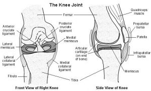
The diagrams below illustrate the knee joint.Each knee joint contains an inner and outer meniscus (medial and lateral meniscus). These are thick rubber-like pads of cartilage tissue. They are C-shaped and become thinner towards the middle of the joint. The meniscal cartilages sit on top of, and are in addition to, the usual thin layer of cartilage which covers the top of the tibia. The menisci act like shock absorbers to absorb the impact of the upper leg on the lower leg. They also help to improve smooth movement and stability of the knee.When people talk about a cartilage injury to a knee, they usually mean an injury to one of the menisci. However, the knee also has cartilage covering the ends of the bones in the joint - this is called articular cartilage - and damage can occur here as well. The areas of articular cartilage can be seen in the side view of the knee joint in the diagram above.What is a sprain?A sprain is an injury to a ligament. Ligaments are strong band-like structures around joints, which attach bones together and give support to joints. A ligament can be injured, usually by being overstretched during a sudden pull. The ligaments at the side of the ankle are the ones most commonly sprained.A damaged ankle ligament causes inflammation, swelling, and bleeding (which shows as bruising) around the affected joint. Moving of the joint is painful. The picture shows a badly sprained ankle with fairly extensive bruising.

The severity of a sprain is graded according to how badly the ligament has been stretched and whether or not the ankle joint has been made unstable. The joint can become unstable when the damaged ligament is no longer able to give it the normal support:
- Grade I - mild stretching of the ligament without joint instability.
- Grade II - partial tear (rupture) of the ligament but without joint instability (or with mild instability).
- Grade III - a severe sprain: complete rupture of the ligament with instability of the joint.
Shin SplintsShin splints is the name often given to exercise-induced pain in the lower leg, specifically along the front of the leg between the knee and the ankle - the area known as the shin. The exact cause of shin splints is not certain but they tend to be as a result of overuse and typically occur in runners. Rest is the most important treatment. Shock-absorbent insoles in your training shoes, graduated running programmes and regularly replacing your training footwear may help in prevention.What are shin splints?

Shin splints is the name often given to exercise-induced pain in the lower leg, specifically along the front of the leg between the knee and the ankle - the area known as the shin.Shin splints are really a symptom rather than a specific diagnosis because they are probably caused by a number of different problems. Shin splints are one of the most common problems in the lower leg in people who exercise or play sports.In typical shin splints, pain is felt more over the inner (medial) part of your shin. Pain felt over the outer (lateral) part of your shin may not be due to shin splints and may be due to a compartment syndrome in your leg.Shin splints are sometimes called medial tibial stress syndrome.Torticollis means 'twisted neck'. The most common cause is acute torticollis, often called 'wry neck'. This is a common cause of neck pain and stiffness. It is common to wake up with a 'wry neck'. It usually goes away on its own over a few days, sometimes longer. Painkillers may ease the pain. Gentle neck exercises are usually advised. There are various other less common causes of torticollis which are briefly discussed below.What is torticollis?Torticollis means 'twisted neck'. The neck becomes twisted to one side. The most common cause of torticollis is acute torticollis, also known as 'wry neck'. Most of this leaflet is about the common acute torticollis. Other less common causes of torticollis are mentioned briefly later in the leaflet.What are the symptoms of 'wry neck' (acute torticollis)?'Acute' means that the symptoms have developed quickly, over a period of hours, or often overnight. The twisting of the neck (torticollis) occurs when the muscles supporting the neck on one side are painful.The cause of acute torticollis is often not known. It can happen in people with no previous neck symptoms. It is a common cause of neck pain in young people. There is usually no obvious injury.However, it may be due to a minor sprain or irritation of a muscle or ligament in the neck. Some reasons for this include:
- Sitting or sleeping in an unusual position without adequate neck support
- Poor posture when looking at a computer screen.
- Carrying heavy unbalanced loads (for example, a briefcase or shopping bag).
- Allowing certain muscles of the neck to be exposed to cold ('sleeping in a draught').
It is common for people to go to bed feeling fine and to wake up the next morning with an acute torticollis.The pain is usually on one side of the neck and stiffness of the muscles in that area twists the neck to one side. Attempts to straighten the neck are difficult due to pain. Occasionally, the pain is in the middle of the neck.The pain may spread to the back of the head or the shoulder. The muscles of the affected side may be tender. Pressure on certain areas may trigger a 'spasm' of the muscle. Movement of the neck is restricted, particularly on one side.
What is a cervicogenic headache?
A cervicogenic headache is simply another name for a headache which originates from the neck and is one of the most common types of headache. It is important to note, however, that there are many types of headache of which cervicogenic is just one. Another common type is vascular (this includes migraines).The spine (neck) comprises of many bones known as vertebrae. Each vertebra connects with the vertebra above and below via two types of joints: the facet joints on either side of the spine and the disc centrally (figure 1).During certain neck movements or sustained postures, stretching or compression force is placed on the joints, muscles, ligaments and nerves of the neck. This may cause damage to these structures if the forces are beyond what the tissues can withstand and can occur traumatically due to a specific incident or gradually over time. When this occurs pain may be referred to the head causing a headache. This condition is known as cervicogenic headache.Cervicogenic headache typically occurs due to damage to one or more joints, muscles, ligaments or nerves of the top 3 vertebra of the neck. The pain associated with this condition is an example of referred pain (i.e. pain arising from a distant source – in this case the neck). This occurs because the nerves that supply the upper neck also supply the skin overlying the head, forehead, jaw line, back of the eyes and ears. As a result, pain arising from structures of the upper neck may refer pain to any of these regions causing a cervicogenic headache.Although cervicogenic headache can occur at any age, it is commonly seen in patients between the ages of 20 and 60.


About 8 in 10 people have one or more bouts of low back pain. In most cases, it is not due to a serious disease or serious back problem, and the exact cause of the pain is not clear. This is called nonspecific lower back pain. The usual advice is to keep active, and do normal activities as much as possible. Painkillers can help until the pain eases. In most cases, the pain disappears within six weeks but may come back (recur) from time to time. Chronic (persistent) pain develops in some cases and further treatment may then be needed.Understanding the lower backThe lower back is also called the lumbosacral area of the back. It is the part of the back between the bottom of the ribs and the top of the legs.Most of the lower back is made up from muscles that attach to, and surround, the spine. The spine is made up of many bones called vertebrae. The vertebrae are roughly circular and between each vertebra is a disc. The discs between the vertebrae are a combination of a strong fibrous outer layer and a softer, gel-like centre. The discs act as shock absorbers and allow the spine to be flexible.

Strong ligaments also attach to nearby (adjacent) vertebrae to give extra support and strength to the spine. The various muscles that are attached to the spine enable the spine to bend and move in various ways.The spinal cord, which contains the nerve pathways to and from the brain, is protected by the spine. Nerves from the spinal cord come out from between the vertebrae to take and receive messages to various parts of the body. Degenerative Disc Disease Degenerative disc disease affects the intervertebral discs that sit between the individual spinal vertebrae. They exist to absorb impact (like a shock absorber) and to prevent bone rubbing on bone. Each disc has a tough outer shell which contains a soft, often described as jelly-like substance, which is primarily made up of water. As the person gets older, the water content decreases and discs can become dehydrated making them more susceptible to injury.The outer layer of the disc can become thinner through wear and tear, or in some circumstances can even tear. These tears can cause the inner substance to leak out into the surrounding structures which may cause pressure to be put on the nerves, alternatively, the nerves may begin to grow in and around the tear of the shell and invade the inner disc space. Either can cause the nerves to become irritated and inflamed. Pain can be chronic with periods of heightened levels, and can usually be helped by keeping mobile so as to relieve pressure from the nerves. Long periods of sitting and standing can aggravate the nerves and increase pain. Lying down can help because the position relieves pressure from the spine.In the event of nerve interference, pain can sometimes be felt down the legs, with numbness or tingling experiences. Giving up smoking is essential if suffering pain from degenerative disc disease. Oxygen levels and blood supply to the area can be compromised with the incidence of smoking. As discs are already at risk of dehydration, reducing the oxygen and nutrients supplied to this area by smoking will exacerbate symptoms further.Osteoarthritis (OA) causes pain and stiffness in joints. Symptoms may be helped by exercises, some physical devices and treatments, and losing weight if you are overweight. Paracetamol will often ease symptoms. Other medicines are sometimes advised. Joint replacement surgery is an option for severe cases.What is osteoarthritis?Arthritis means inflammation of the joints. Osteoarthritis (OA) is the most common form of arthritis in the UK. OA mainly affects the joint cartilage and the bone tissue next to the cartilage.Understanding joints

A joint is the term for where two bones meet. Joints allow movement and flexibility of various parts of the body. The movement of the bones is caused by muscles which pull on tendons that are attached to bone.Cartilage is a hard, smooth tissue that covers the end of bones. Between the cartilages of two bones which form a joint, there is a small amount of thick fluid called synovial fluid. This fluid lubricates the joint, which allows smooth movement between the bones.The synovial fluid is made by the synovium. This is the tissue that surrounds the joint. The outer part of the synovium is called the capsule. This is tough and helps to give the joint stability. Surrounding ligaments and muscles also help to give support and stability to joints.